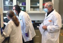
Just a block or so from the busy shops and restaurants of Providence’s Thayer Street, a potentially significant research project is underway that requires soft lights, whispering research assistants and a quiet baby nursery.
Nearly every night of the week, babies of all shapes and sizes visit Brown University’s Advanced Baby Imaging Lab to take part in a groundbreaking study that uses magnetic resonance imaging – a procedure normally reserved for older youngsters and adults – to explore infant brain development.
The lab was created five years ago with a $2.4 million grant from the National Institute of Mental Health after Sean C. Deoni, an assistant professor of engineering at Brown, figured out a way to take an MRI of an infant without using sedation. The result is a place that is fun and serious at the same time, where teddy bears are abundant and cartoon figures adorn the walls, but research is being conducted that may one day lead to greater understanding of autism, attention deficit and other neurological problems.
“This was something we needed,” Judith S. Mercer, a nursing professor at the University of Rhode Island, said of the lab.
Mercer and URI colleague Deb Erickson-Owens are using the lab to study the role increased iron – due to delayed clamping of the umbilical cord – plays in the creation of myelin, a fatty substance that helps to speed up brain messages. In 2012, the researchers received a $2.4 million, five-year grant from the National Institutes of Health for their study, which they believe they would not have received without the already online Advanced Baby Imaging Lab.
The lab is the sort of innovative facility that Rhode Island wants to encourage as it strives to create a knowledge-based economy. It is attracting attention from researchers in Europe and California, as well as from the University of Rhode Island, who see potential in the techniques Deoni has developed for infant brain imaging.
“[Deoni] is the first in the world to examine newborn brain development using an MRI,” Mercer has said.
Hospitals, including Hasbro Children’s Hospital, sometimes take MRIs of babies for diagnostic purposes, but they usually sedate them first to stop them from squirming.
Deoni’s method, which he developed in England before joining Brown’s faculty in 2009, is long on common sense, aided by a few simple devices.
First, the MRIs are scheduled only at night, when the baby presumably wants to sleep. Secondly, the crib used in the lab’s makeshift nursery is outfitted with an underlying pallet that enables researchers to transfer the child by lifting the pallet and not the baby, reducing the chances of waking the infant. Finally, the baby sleeps on a special inflatable mattress that, once blown up, conforms and hardens to the child’s shape, holding the baby still.
Tiny ear plugs are also used to reduce the noise during the 30-minute exam, half the length of time for a normal MRI.
Sometimes, despite these measures, a baby will wake up during the MRI. When this happens, Deoni and his assistants must do what every parent tries to do in the same situation – lull the child back to sleep.
“It’s a great deal of fun,” Deoni said of his work with the babies.
When his five-year study is completed later this year, he will have taken brain images of 410 infants and toddlers, some of them three or four times over a period of two years, creating an invaluable set of normal infant brain pictures. Having a true picture of normal infant brain development is crucial to understanding what’s abnormal, he and others said.
The data has already yielded evidence of the value of breast feeding to infant brain development, but it could hold the answers to many more scientific questions, according to Deoni. He called it a “treasure trove” of information.
The imaging takes place on the ground floor of the Sidney E. Frank Hall for Life Sciences at Brown, where each night Deoni and his team convert a small, empty room adjacent to the MRI machine into a cozy nursery. Across the hall, three additional rooms are used as play areas for waiting children and their parents.
In addition to finding a way to keep the babies still, Deoni has developed an imaging technique for capturing the myelination process in the infant brain. His work caught the attention of Mercer and Erickson-Owens, after they heard him speak at Women & Infants Hospital in early 2010, resulting in their collaborative research on the effects of delayed cord-clamping on myelin development in full-term babies.
In a prior study, Mercer looked at the effect of delayed cord clamping on premature babies. This research, which received a $100,000 seed grant from the Bill & Melinda Gates Foundation in addition to federal funding, found that even delaying the clamping by just 30 to 45 seconds increased the flow of iron-rich blood to the baby, which in turn has been linked to several benefits.
For the new study, which is now in its second year, the researchers want to measure the effect of a five-minute delay in clamping on full-term babies. Their study involves taking MRIs of 128 healthy babies over a three-year period at the Advanced Baby Imaging Lab.
“We have wanted to look at full-term babies for several years,” but didn’t know how, said Mercer, until now with the help of the Advanced Baby Imaging Lab. •
No posts to display
Sign in
Welcome! Log into your account
Forgot your password? Get help
Privacy Policy
Password recovery
Recover your password
A password will be e-mailed to you.


