
When doctors want to know how well your heart is pumping, how the blood is flowing, where vessels are obstructed, the best way to do that is to inject a tiny amount of radioactive materials and watch as they move through the system.
Each year, millions of Americans undergo nuclear cardiology studies, during exercise and at rest. In addition, nuclear medicine is used to diagnose or exclude diseases in every major organ system, often in earlier stages than with any other method. It’s even used to treat hyperthyroidism and thyroid cancer, and to ease the pain from bone metastases.
Now The Miriam Hospital’s nuclear medicine department, which performs more than 6,000 studies per year, has earned two national accreditations for its programs, both in general nuclear medicine and nuclear cardiology, from the Intersocietal Commission for the Accreditation of Nuclear Medicine Laboratories.
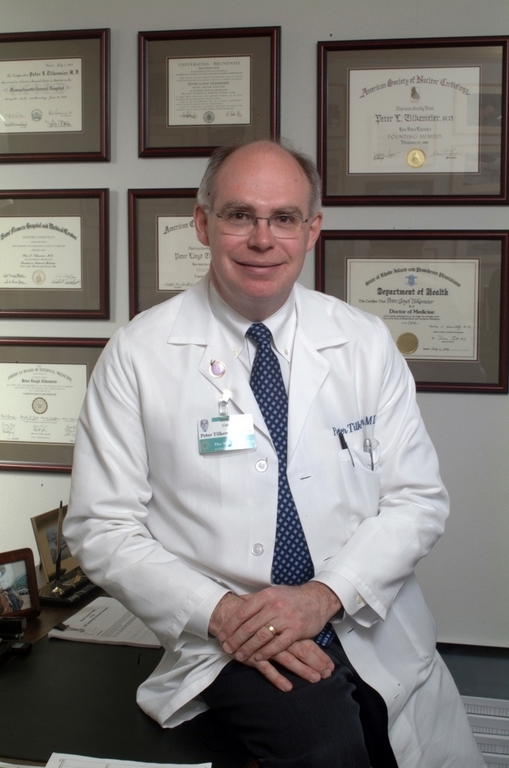
Dr. Peter Tilkemeier, a veteran nuclear cardiologist at the Miriam and an associate professor at The Warren Alpert Medical School of Brown University, explained how nuclear medicine works and why getting accredited was important to the hospital.
PBN: How does nuclear medicine work, and what are its main uses?
TILKEMEIER: The difference with nuclear medicine compared with most other imaging techniques is that it allows us to see the actual function of an organ or a cell or a body part, as opposed to looking at its structure, as you might with a CT scan.
When we give a patient a radioactive substance, it is directed toward answering a specific question. So I’m a nuclear cardiologist; if you come to me with chest pain and I want to see blood flow to your heart muscle, we use a specific radioactive pharmaceutical that is supposed to go to the heart muscle, and if it doesn’t go there, we know there’s a problem with the blood flow that will allow us to diagnose coronary artery disease.
Nuclear medicine can also be applied to lungs, livers, bones, brains, thyroids – it’s a generalized field that we use to image organ function throughout the body, using small amounts of radioactivity that are tagged to go to certain body organs.
Seifert Systems Invests in Energy Efficiency to Strengthen Operations
For manufacturers, energy is more than just another operating expense. It plays a critical role…
Learn More
PBN: How is it tagged?
TILKEMEIER: We couple it to a chemical that is usually part of the metabolism of that organ, so if we’re tagging the thyroid, for example, we’ll couple that with iodine, because iodine is taken up by the thyroid. For cardiac imaging, we couple it to a chemical substance that the heart muscle cells need for them to function. So it drags the radioactivity along with the substance that the organ we need to study uses.
PBN: How tightly are you regulated in this work?
TILKEMEIER: We are very heavily regulated, at the state level and at the federal level, for the safe operation and handling of radiation. The accreditation process is voluntary, part of a quality program nationally to recognize laboratories that have achieved a high level in patient care, imaging, quality assurance, reporting and communication of the results to the referring physician. There are two aspects of accreditation, cardiac and general nuclear medicine. This is our third three-year cycle that we’re accredited for on the cardiac side; this is our second cycle for which we’ve been accredited for general nuclear medicine.
Now, it may seem simple, but the standards are constantly changing as new things are developed in the field, and you have to keep up. There are only 100 labs in the entire United States that are accredited for nuclear medicine, and only one in Rhode Island, at the Miriam. For cardiology, there are larger numbers.
PBN: There’s been such an explosion of new imaging technologies in the last few years. How does nuclear medicine stack up against that? Is there anything that’s doing the job better, or is this still the best we have?
TILKEMEIER: PET – positron emission tomography – imaging is now a sub-area of nuclear medicine, because we’re using the nuclear medicine isotopes, and that’s probably the newest technology we have in the field of nuclear medicine. But there’s nothing else, really. The other technologies that are out there are looking at structural information of organs, and they can do so in exquisite detail, but there’s nothing that’s looking at how that cell is functioning, how it’s interacting with the chemicals and the radioactive materials that we give it, and how that’s going to affect the patient’s outcome. And that’s nuclear medicine.
PBN: Is your field actually growing?
TILKEMEIER: I would say we’re stable. We’re working with the other imaging modalities – magnetic resonance, CT imaging – to find out how we could use different modalities in concert with one another to give you better diagnostic information. So, for example, doing a CT scan at the same time as your nuclear medicine study, so we can layer the structural information with the functional information, which would give us a much more powerful diagnostic technique.
The Miriam Hospital is a member of the Lifespan health system and is affiliated with The Warren Alpert Medical School of Brown University. For more information, go to www.miriamhospital.org.